Please visit the Duke Health site to learn about the various types of cancer we treat.
We are proud of our partnerships and expansions. Read about our affiliated health centers here. Please note that treatment options at these centers may vary.
We offer a full range of radiation therapy treatments:
Conformal radiation therapy utilizes imaging and computer software tools to help the physician better design radiation treatment beams. This procedure involves the construction of an immobilization device to be used during treatment planning and treatment. Three-dimensional imaging (such as computed tomography) is done with the patient in the immobilization device. The physician then plans treatment beams on the images. Once appropriate beams are designed, the patient returns to the radiation department (generally one week following the initial scan) for a confirmation "physical simulation," followed by the initiation of treatment.
This therapy is most often used to treat primary choroidal melanoma and retinoblastoma. We construct an eye plaque made out of gold, which holds radioactive Iodine-125 seeds. Each plaque is custom made to provide individualized treatment for each tumor. At the time of surgery, the plaque is positioned on the outside of the eye against the base of the tumor. The plaque emits radiation over 5 days to kill the tumor cells so that the patient’s eye can be preserved. The eye plaque is removed after treatment.
Duke University Medical Center was one of the founding institutions of the Collaborative Ocular Melanoma Study, designed to evaluate the efficacy of eye plaque therapy. We have extensive experience with Iodine-125 plaque therapy for choroidal melanomas and preoperative irradiation for tumors that are too large for eye plaques. The Department also has extensive experience in the treatment of retinoblastoma.
Brachytherapy entails the placement of a radioactive source in a cavity (intracavitary therapy) in close proximity to the tumor or in the tissues themselves (interstitial therapy) to deliver a high dose of radiation to a limited volume. High Dose Rate (HDR) brachytherapy involves using a radioactive source, such as iridium, to deliver a high dose of radiation in a very short period of time. The HDR unit is small, self-contained and can be moved about. While the unit is not in use, the radioactive source is in the safe and poses no risk of radiation exposure.
During a procedure the radioactive source is out of the safe and there is the risk of radiation exposure to health care personnel. To avoid this, the procedures are often carried out with the patient in an existing, shielded accelerator room with the health care personnel outside of the room. At the time of surgery catheters can be placed in the area of the tumor and – with the patient still under anesthesia – the patient is brought to the booth adjacent to the operating room. With the catheters in place, the radioactive source is passed through the catheters for the specified time necessary to deliver the desired dose of radiation. This unit can also be used for some of the cases currently being done with conventional, LDR radioactive sources. HDR does not replace LDR brachytherapy; it expands this type of resource.
LDR refers the use of inserted or implanted radioactive sources for various cancers, such as the treatment of cervical cancers using intracavitary implantation of Cs-137 brachytherapy sources, PSI, eye plaque treatment and more.
The intraoperative radiation therapy program is a joint effort of the Departments of Radiation Oncology, Surgery, Gynecologic Oncology and Anesthesiology. In this highly specialized technique, radiation therapy is administered via a high dose rate afterloading machine during surgery.
Treatment to tumors located in the thoracic and abdominal regions, such as lung and liver, is challenging because of respiratory organ motions. The traditional approach is to expand each treatment field to ensure the entire tumor volume is included in the radiation field. To minimize radiation damage to extra normal tissues due field expansion, Duke has adopted respiratory control techniques to synchronize the radiation beam with tumor motion. With this technique, the treatment margin added to accommodate the organ motion could be reduced to minimize the radiation dose to normal tissues. Advanced technologies are used to achieve this goal, such as the use of Varian RPM system to monitor respiratory pattern, On-Board Imager to verify target motion, and electronic portal imaging system to verify real-time radiation delivery.
Image-Guided Radiation Therapy (IGRT) is a process of using various imaging technologies to locate a tumor target prior to a radiation therapy treatment. This process is aimed to improve treatment accuracy so that the need for wide target margins, which have traditionally been used to compensate for errors in localization. As a result, the amount of healthy tissue exposed to radiation can be reduced, minimizing the incidence of side effects. At Duke, physicians applied advanced imaging techniques using CT, MRI, PET/CT and SPECT to accurately delineate treatment target. Moreover, they are able to use state-of-art in-room on-board imaging and cone-beam CT technologies to visualize the treatment tumor three-dimensionally prior to the delivery of radiation beam. IGRT is complementary to IMRT. IMRT is used to improve the radiation delivery precision and IGRT is used to improve the radiation delivery accuracy.
Tumors to be treated are often surrounded by normal tissue and/or organs. The dose tolerances to the normal critical organs are one of the limiting factors to radiation dose prescribed to tumor. With the IMRT technique, one will be able to shape the radiation dose to the target and then minimize the dose to critical organs. Therefore, the expected dose could be prescribed to tumor while limiting the radiation damage to the normal tissue within the tolerance. The IMRT involves a complicated treatment planning process (called inverse planning) and a dedicated delivery process using dynamic multi-leaf collimator (MLC). A comprehensive quality assurance program specific for both machine and patient was very critical and was developed at Duke to ensure the quality treatment of IMRT. We use the latest Varian Eclipse inverse planning system and the Clinac 21EX machine with120-leaf MLC to deliver fine radiation dose to the target.
Prostate Seed Implant (PSI) is a treatment procedure for the use of permanently implanted radioactive sources for treatment of prostate cancer. This procedure was conducted with team efforts from the Departments of Radiation Oncology and Urology. It is also one of low-dose rate brachytherapy procedures.
Stereotactic Radiosurgery (SRS) is a radiation technique that uses precise patient positioning and extremely accurate radiation delivery to treat brain lesions in one or up to five treatments. At Duke, SRS is delivered by a Novalis TX or Varian TrueBeam STX linear accelerator. These machines have state-of-the-art technology, which allows your radiation oncologist to give treatment with great precision, speed and patient comfort. SRS is used to treat many types of brain tumors. The use of SRS is limited by the size of tumors, which generally need to be less than 5 cm in maximum diameter (about 2 inches). Larger lesions up to approximately 3 cm (about 1.25 inches diameter) can often be treated in a single session, while larger tumors may require five, lower dose treatments. Treatable tumor types include metastatic lesions (meaning a brain tumor which develops as the result of spread from another primary site), such as breast, lung, melanoma or renal cell cancer. More recently, SRS has been used to treat malignant gliomas, such as glioblastoma (GBM) and anaplastic astrocytoma, that have recurred after previous treatment with surgery, radiation therapy and chemotherapy. SRS can also effectively treat small, non-malignant tumors, such as meningiomas, acoustic neuromas (also known as vestibular schwannomas) and glomus tumors. SRS is used to treat other brain abnormalities, including arteriovenous malformation (AVM) and trigeminal neuralgia (severe facial pain originating from the trigeminal nerve).
Stereotactic Body Radiotherapy (SBRT) uses the same precise techniques to treat tumors outside of the brain. This is most often used for small tumors in the lung, liver or spine. SBRT is used to treat metastatic tumors in the spine, lung, liver, abdominal or pelvis areas. There are limitations to using SBRT including the size and number of lesions. Early stage lung cancer can be treated with SBRT alone instead of undergoing lung surgery. Most often SBRT requires 3-5 treatments, but can be completed in one session if the lesion is small enough.
All radiosurgery procedures are performed by a multidisciplinary team consisting of a Radiation Oncologist, radiation physicist, radiation therapists, radiosurgery-specific treatment planners and, depending on the site of the body to be treated, a neurosurgeon, neurotologist or thoracic oncologist. Duke physicians, physicists, planners and therapists are actively involved in radiosurgery clinical trials, teaching, technology assessment and development of quality assurance systems.
WHAT IS THE BENEFIT?
At the time of the consultation visit with your radiation oncologist, the specific benefits of SRS vs. any other treatment options will be discussed in detail. In general, SRS for metastatic lesions is very well tolerated with few expected side effects. Since SRS can be completed in a short period of time, it allows the patient to move on quickly to other recommended treatments such as surgery or chemotherapy. For recurrent gliomas, the best option for safely delivering a second course of radiation therapy is using this highly precise SRS technique.
HOW OFTEN DOES A PATIENT NEED TREATMENT?
SRS can be completed in a relatively short timeframe. Every patient will have an initial consultation visit with a radiation oncologist specializing in their diagnosis. Once SRS is agreed upon, a customized plastic-mesh face mask will be made, which will maintain your head in the correct position for treatment planning and subsequent delivery. A CT scan is obtained while you are wearing the mask, which will be used to accurately position your head during the SRS treatment, as well as a new MRI scan. The SRS treatment is usually ready to be performed in about 4 working days after the planning procedures are done. For larger tumors, SRS may need to be done daily for 5 treatments.
SRS for treatment of trigeminal neuralgia requires a different method of head immobilization – a lightweight metal stereotactic frame. The frame is placed and is followed shortly by a CT scan. An MRI scan is also used to plan the treatment, but this scan is performed one or more days before the day of SRS. The SRS procedure requires careful and precise planning and generally is ready to be performed by early afternoon. The head frame is removed as soon as the SRS procedure is done.
SBRT requires another type of immobilization device. This device would either be a foam-based mold or a cradle made via a vacuum. A CT scan is also performed while the patient is in the treatment device to begin the radiation planning. The first treatment will be scheduled in approximately one week.
HOW LONG DOES THE TREATMENT LAST?
SRS and SBRT treatments are outpatient procedures that take about 45 minutes to complete for one lesion, and somewhat longer for more than one lesion. The majority of the treatment time involves assuring your position is perfectly aligned before each x-ray beam is given. Your radiation therapist will keep you informed about your progress throughout the treatment process. Discharge instructions will be given to you at the SRS completion, including when to return for a checkup and MRI scan.
WHAT ARE THE SIDE EFFECTS?
Side effects from a SRS or SBRT procedure vary depending on the location, size and number of lesions that are treated. During the consultation appointment, any potential side effects will be discussed in detail with you by your radiation oncologist.
Total Body Irradiation (TBI) is radiation given to the whole body as a preparatory regimen for bone marrow transplant in the treatment of hematologic cancers and other disorders.
Total Skin Electron Beam (TSEB) is radiation given to all of the skin with an electron beam technique for various skin cancers.
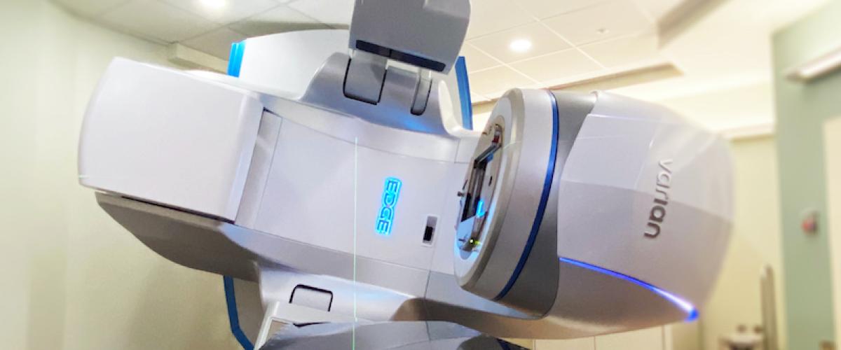
Technology
Duke Radiation Oncology treats with a complete range of radiation therapy equipment, including simulators (with CT capability), linear accelerators, two- and three-dimensional treatment planning computers, a high dose brachytherapy unit and a dedicated intraoperative radiation therapy program. At the Duke Cancer Center, our machines are listed by color as an internal way for our staff to identify machines.
Learn about our machines at the Duke Cancer Center:
- Truebeam STX
- 6/10/15 MV photons, 6/10 MV Flattening filter free photons and 6/9/12/16/20 MEV electrons
- MV and KV 2D imaging and KV 3D imaging (CBCT)
- Motion management
- Treatment: electron/2D/3D/IMRT/SBRT
- Red machine is capable for TBI
- Truebeam STX
- 6/10/15 MV photons, 6/10 MV flattening filter free photons and 6/9/12/16/20 MEV electrons
- MV (portal vision) and KV
- Motion management
- Exactrac system with 6 degree couch
- High definition MLC
- Treatment: electron/2D/3D/IMRT/TSI/SBRT/SRS
- Varian 21EX
- 6/15 MV photons and 6/9/12/16/20 MeV electrons
- MV (portal vision) and KV (OBI) 2D imaging and KV 3D imaging (CBCT)
- Motion management using RPM
- Treatment: electron/2D/3D/IMRT
- Varian 600CD
- 6 MV photon
- MV (portal vision) 2D imaging
- Treatment: 2D/3D/IMRT
- Varian GammaMedplus IX remote afterloader
- 24 channels
- 0.1 seconds dwell time resolution
- 1 to 10 mm step sizes
- Console computer system